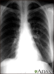
Coccidioidomycosis - chest x-ray

This chest x-ray shows the affects of a fungal infection, coccidioidomycosis. In the middle of the left lung, there are multiple, thin-walled cavities (seen as light areas) with a diameter of 2 to 4 centimeters. To the side of these light areas are patchy light areas with irregular and poorly defined borders.
Other diseases that may explain these x-ray findings include lung abscesses, chronic pulmonary tuberculosis, chronic pulmonary histoplasmosis, and others.

|
Review Date:
5/21/2012 Reviewed By: Harvey Simon, MD, Editor-in-Chief, Associate Professor of Medicine, Harvard Medical School; Physician, Massachusetts General Hospital. |
The information provided herein should not be used during any medical emergency or for the diagnosis or treatment of any medical condition. A licensed medical professional should be consulted for diagnosis and treatment of any and all medical conditions. Links to other sites are provided for information only -- they do not constitute endorsements of those other sites. © 1997-
A.D.A.M., Inc. Any duplication or distribution of the information contained herein is strictly prohibited.

